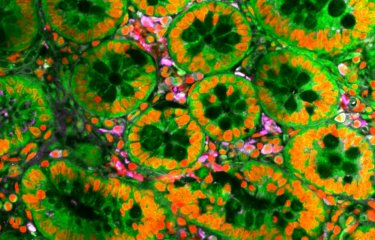
SODA (Statistical Object Distance Analysis) is a new method for performing automatic statistical analysis on images and determining the spatial distribution of molecules. It has been made available free of charge for the scientific community via Icy and does not require any programming knowledge. This software is applicable to all fluorescence imaging and can be used for either conventional imaging or super-resolution imaging (SIM, STED or STORM).
"To establish the precise localization of the various molecular players in the cell and understand how they are organized and how they function, we need to carry out a statistical analysis of their spatial distribution using high-resolution microscopy images," explains Thibault Lagache, a post-doctoral fellow in the Bioimage Analysis Unit (Institut Pasteur/CNRS UMR3691) directed by Jean-Christophe Olivo-Marin.
The scientist co-authored a paper published recently in Nature Communications describing how the use of a novel algorithm for statistical image analysis provided new information about the organization of proteins at the synapse of hippocampal neurons.
This algorithm, dubbed SODA (Statistical Object Distance Analysis), enables the statistical identification of coupled molecules and the production of spatial maps showing coupled or isolated molecules. In conventional microscopy, scientists previously referred to "colocalizations" when describing spatial overlap in the clusters observed. Since the emergence of new super-resolution microscopy techniques, which offer significant improvements in resolution, the term "coupling" has instead been employed to describe molecules that are found together but do not necessarily have the same spatial coordinates.
SODA was developed by Thibault Lagache during his post-doctoral fellowship in the Bioimage Analysis Unit. It enables scientists not only to analyze colocalizations/coupling between molecules imaged using conventional and super-resolution microscopy, but also to generate spatial maps of the molecule pairs or triplets analyzed.
The paper explains how SODA was applied to images obtained via three-color structured illumination microscopy (SIM) to describe the spatial organization of thousands of synapses, in collaboration with Lydia Danglot, Scientific Coordinator of the NeurImag imaging platform at the new Paris Center for Psychiatry and Neuroscience (Unit 894 Inserm/Paris Descartes University, Paris). This technique revealed the asymmetric organization of the proteins Synapsin, Homer and PSD95. Used in conjunction with 3D STORM technology, it determined the relationships between thousands of single molecule localizations within synaptic boutons. In collaboration with the group led by Nathalie Sauvonnet (Molecular Microbial Pathogenesis, Institut Pasteur/Inserm U1202), SODA was also used to study different endocytic pathways.
To encourage the use of SODA by the broader scientific community and facilitate reproducibility of results, the tool has been made available free of charge via the platform Icy.
Find out more about the potential of SODA and SODA STORM
For more information, please visit


