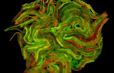
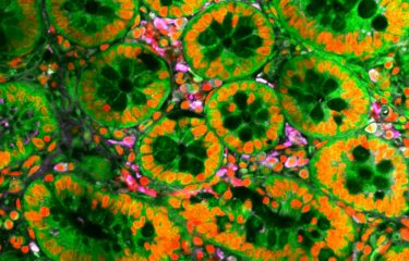
Biological image analysis using mathematical methods, artificial intelligence and computer visualization techniques has become an essential tool for biological research and an effective driver for new discoveries. In a publication in Patterns (Cell Press)1, scientists from the "Bioimage Analysis Unit" recently presented the results of multidisciplinary research using quantitative image analysis to shed light on cell motility.
Institut Pasteur scientists have put forward a robust process that includes all tasks required for performing an exhaustive analysis of moving cell images, and understanding inherently non-linear, multi-factorial phenomena.
A major challenge for research
In biology, images or signals from microscopes are digitized for computer processing, with a view to quantitatively characterizing studied phenomena. This article looks at how the quantitative analysis of images from moving cells has contributed to knowledge of invasive cellular processes. The scientists are chiefly interested in cell movement and migration, for which the latest data have led to the implementation of complex experimental systems aimed at reproducing physiologically relevant conditions. This is a major challenge when determining how pathogenic organisms (viruses, bacteria, and parasites) penetrate, move within, and invade human tissue.
Diverse quantification strategies are required due to the diversity of cell motility
When dissecting the molecular machinery underpinning cell movement, research is impacted by a combination of at least three theoretical and experimental processes: microscope imaging, the laws of physics, and spatial architecture combined with the chemical nature of the extracellular environment. Quantitative image processing helps increase the objectivity and reproducibility of this eminently qualitative science. However, no current mathematical model incorporates the diverse range of biological mechanisms required to fully predict cell migration.
Detection, characterization, and monitoring of cells in microscopic imaging
The experiments conducted by the authors evolved over time, closely mirroring developments in image processing techniques. Algorithms have been developed to detect and segment cells as independent objects, temporally monitor their movement, and define morphological descriptors.
While studying amoeboid movement as a factor guiding intestinal infection by pathogenic amoebae, these image analysis methods have helped illustrate how appropriate quantification of cell behavior provides insights on biological data, e.g. by choosing mathematical representations that amplify initially subtle differences, statistically determining general laws governing cell movement in two or three dimensions and in confined environments (such as tissue), and incorporating a physical view of phenomena2,3. In the current research, the authors reveal that quantitative imaging is non-invasive, providing a further boost to two flourishing fields. The first is mechanobiology, for which many biophysical measurements are still impossible. The second is the fabrication of 3D microenvironments, in which efforts to achieve physiological relevance for selected phenomena have led to a significant increase in volumes of computer data.
Sources
[1]. Bioimage Analysis and Cell Motility. Patterns, Volume 2, Issue 1, 2021
Aleix Boquet-Pujadas,1,2,3 Jean-Christophe Olivo-Marin,1,2,* and Nancy Guillen 1,4
1Institut Pasteur, Bioimage Analysis Unit, 25 rue du Dr. Roux, Paris Cedex 15 75724, France
2French National Center for Scientific Research, CNRS UMR3691, Paris, France
3Sorbonne University, Paris 75005, France
4French National Center for Scientific Research, CNRS ERL9195, Paris, France
*Correspondence: jcolivo@pasteur.fr
https://doi.org/10.1016/j.patter.2020.100170
[2] BioFlow: a non-invasive, image-based method to measure speed, pressure and forces inside living cells. Sci Rep. 2017 7(1):9178, Boquet-Pujadas A1,2,7, Lecomte T1,2,7, Manich M 1,2, Thibeaux R 3,4,5, Labruyère E 1,2, Guillén N 3,4,6, Olivo-Marin JC 11,2, Dufour AC 1,2
1 Institut Pasteur, Bioimage Analysis Unit, Paris, France
2 CNRS UMR3691, Paris, France
3 Institut Pasteur, Cell Biology of Parasitism Unit, Paris, France
4 INSERM U786, Paris, France
5 Institut Pasteur, Leptospirosis Research Unit, New Caledonia
6 CNRS ERL9195, Paris, France
7Aleix Boquet-Pujadas and Timothée Lecomte contributed equally to this work.
Corresponding author: Jean-Christophe Olivo-Marin
email: jcolivo@pasteur.fr
[3] E-cadherin focuses protrusion formation at the front of migrating cells by impeding actin flow. Nature Communications volume 11, 2020 Cecilia Grimaldi, Isabel Schumacher, Aleix Boquet-Pujadas, Katsiaryna Tarbashevich, Bart Eduard Vos, Jan Bandemer, Jan Schick, Anne Aalto, Jean-Christophe Olivo-Marin, Timo Betz & Erez Raz
https://doi.org/10.1038/s41467-020-19114-z


