The human body is made up of tens of thousands of billions of cells, all originating from the division of one parent cell into two daughter cells. Scientists from the Institut Pasteur set out to investigate midbodies, which are key cell division structures, and they identified the role of BST2, a restriction factor involved in viral infections.
In order to divide, cells start by making identical copies of their chromosomes before distributing them to daughter cells. The parent cell then needs to physically cut itself into two daughter cells in a complex process known as 'cytokinesis'. Defects of cytokinesis result in the formation of genetically unstable cells, which are thought to account for over 40% of human cancers.
'Midbodies': key structures in cytokinesis
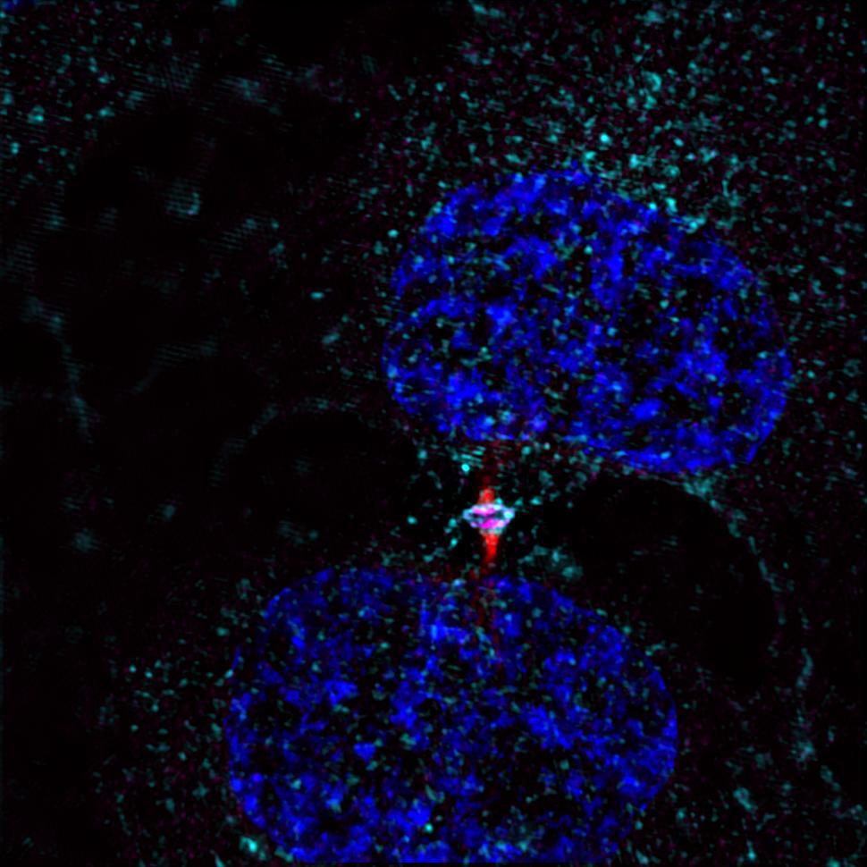
Cytokinesis begins when a parent cell contracts at its center, resulting in two daughter cells connected by an intercellular bridge. At the center of this bridge, there is a specific structure known as a 'midbody', which comprises all of the proteins needed to finally cut the parent cell into two daughter cells. Following this abscission, which actually takes place either side of the midbody, a ‘midbody remnant' (MBR) is released outside the cells. Intensive research is currently being conducted on this MBR, because it seems to contain information that influences the proliferation and differentiation of the cells which capture it. However, we know little about the mechanisms which allow the MBR to be captured or retained by the target cells.
Discovery of the BST2 protein's involvement in midbodies
The Membrane Traffic and Cell Division Unit (Institut Pasteur - CNRS, directed by Arnaud Echard) in the Department of Cell Biology and Infection is focusing on the molecular mechanisms associated with cytokinesis in human cells. Recently, this team produced an inventory of the proteins present in the midbody prior to abscission. Tetherin/BST2 was unexpectedly revealed to be one of these proteins. BST2 is well known to virologists as a restriction factor that retains various enveloped viruses (including HIV-1, the retrovirus that causes AIDS) at the surface of infected cells, thus restricting their spread within the body.

Midbody in a bridge (in red), BST2 in cyan blue during cytokinesis, before abscission. Credit: Adrien Presle, Audrey Salles, Arnaud Echard/Institut Pasteur
A parallel between the role of BST2 in viral infections and in the capture of midbodies
Scientists from this research unit have now shown that, in a similar way, this same protein helps to retain and capture the MBRs generated during cell division. As seen in viruses, a lack of BST2 promotes the release of MBRs into the extracellular space as well as their spread over long distances and restricts their capture by target cells.
This research thus reveals the key role played by BST2 in non-infected cells. It also highlights a surprising parallel between viruses and a structure that is generated naturally during cell division. BST2 is frequently overexpressed in human tumors, which could potentially promote the capture of MBRs and thus contribute towards cell proliferation.
Source:
The Viral Restriction Factor Tetherin / BST2 Tethers Cytokinetic Midbody Remnants to the Cell Surface, Current Biology, March 11, 2021
Adrien Presle1,2, Stéphane Frémont1, Audrey Salles3, Pierre-Henri Commere4, Nathalie Sassoon1, Clarisse Berlioz-Torrent5, Neetu Gupta-Rossi1,6 and Arnaud Echard1,6
1 Membrane Traffic and Cell Division Unit, Institut Pasteur, UMR3691, CNRS, 25-28 rue du Dr Roux, F-75015 Paris, France
2 Sorbonne University, Doctoral school, F-75005 Paris, France
3 UTechS Photonic BioImaging PBI (Imagopole), Center for Technological Resources and Research C2RT, Institut Pasteur, F-75015 Paris, France
4 UTechS CB, Center for Technological Resources and Research C2RT, Institut Pasteur, F-75015 Paris, France
5 Université de Paris, Institut Cochin, INSERM, CNRS, F-75014 Paris, France
6These authors contributed equally


