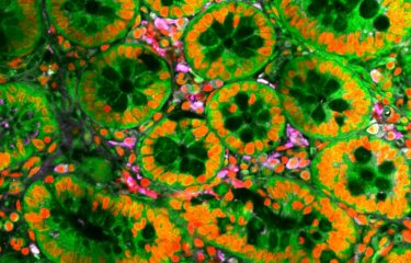
Cardiovascular diseases are a major cause of mortality and morbidity worldwide. After myocardial infarction, heart cells (or cardiomyocytes) die and are replaced by fibrotic tissue leading to impaired cardiac function. The assessment of the regenerative capacity of the heart has always been compromised by the lack of signatures to characterize cardiomyocytes. Researchers from the Institut Pasteur discovered a population of immature cardiomyocytes which persists in the adult heart and is able to respond to infarction by increasing in numbers. This work opens new perspectives in the understanding and treatment of heart pathologies.
Cardiomyocytes contribute to heart growth through extensive cell divisions and exit the cell cycle as development progresses, a process completed by the end of the first week of postnatal
life. By combining multiparametric surface marker analysis with single cell transcriptional profiling and in vivo transplantation, researchers identified the main mouse embryonic cardiac populations, allowing tracing the main cardiac lineages from their developmental origins.
Researchers identified a population of immature cardiomyocytes that are present throughout life. This immature subset that co-exists with mature cardiomyocytes actively proliferated in vivo up to day 7 post-birth. “These immature cardiomyocytes that persist in small proportions in the adult heart respond to myocardial infarction by proliferating, albeit at insufficient rates to ensure regeneration of the myocardium” explains Ana Cumano, head of the Lymphopoiesis Unit (Pathophysiology of the immune system, Inserm U1223), at the Institut Pasteur. This work however found no evidence for the persistence of a self-renewing compartment of cardiomyocyte stem cells in the adult heart.
“The possibility to isolate immature cardiomyocytes poised for activation in response to infarction at any developmental stage opens new perspectives in the understanding of heart pathologies and is of interest to the fields of molecular development and regenerative medicine” she concludes.
This work that has been financed through the LabEx REVIVE (Investissement d’Avenir; ANR-10-LABX-73) illustrates how studies aiming at identifying and isolating the cells that participate to cardiac muscle formation, during embryonic development, can help elaborating strategies for adult heart repair.
Source
Mouse HSA+ immature cardiomyocytes persist in the adult heart and expand after ischemic injury, PLOS Biology, July 10th, 2019
Mariana Valente1,2,3,4, Tatiana Pinho Resende1,2, Diana Santos Nascimento1,2, Odile Burlen-Defranoux4,5, Francisca Soares-da-Silva1,2,3,4, Benoit Dupont6, Ana Cumano4,5, Perpetua Pinto-do-O1,2,3,4
1 Instituto de Investigacão e Inovacão em Saude (i3S), Universidade do Porto, Porto, Portugal
2 Instituto Nacional de Engenharia Biomedica (INEB), Universidade do Porto, Porto, Portugal
3 Instituto de Ciências Biome´dicas Abel Salazar (ICBAS), Universidade do Porto, Porto, Portugal
4 Unit for Lymphopoiesis,Immunology Department, INSERM U1223, Institut Pasteur, Paris, France
5 Université Paris Diderot, Sorbonne Paris Cité , Cellule Pasteur, Paris, France
6 Beckman Coulter, France S.A.S, Villepinte, France


