Researchers from the Institut Pasteur and CNRS have set up a new optical microscopy approach that combines two recent imaging techniques in order to visualize molecular assemblies without affecting their biological functions, at a resolution 10 times better than that of traditional microscopes. Using this approach, they were able to observe the AIDS virus and its capsids (containing the HIV genome) within cells at a scale of 30 nanometres, for the first time with light. This newly developed approach represents a significant advance in molecular biology, opening the door to less invasive and more precise analyses of pathogenic microorganisms present in human host cells. This study is already published in the Electronic Edition of PNAS.
Press release
Paris, may 16, 2013

A study coordinated by Dr Christophe Zimmer(1) (Institut Pasteur/CNRS), in collaboration with Dr Nathalie Arhel(2) within the lab headed by Pr Pierre Charneau(3) (Institut Pasteur/CNRS), shows that the association of two recent imaging techniques helps obtain unique images of molecular assemblies of HIV-1 capsids, with a resolution around 10 times better than that of traditional microscopes. This new approach, which uses super-resolution imaging and FlAsH labeling, does not affect the virus’ ability to self-replicate. It represents a major step forward in molecular biology studies, enabling the visualisation of microbial complexes at a scale of 30 nm without affecting their function.
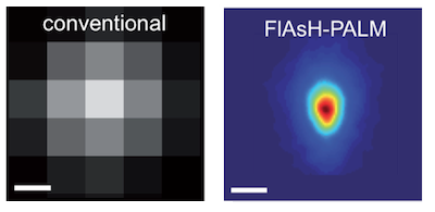
The newly developed approach combines super-resolution PALM imaging and fluorescent FlAsH labeling. PALM imaging relies on the acquisition of thousands of low-resolution images, each of which showing only a few fluorescent molecules. The molecular positions are then calculated with high accuracy by computer programs and compiled into a single high-resolution image. FlAsH labeling involves the insertion of a 6-amino-acid peptide into the protein of interest. The binding of the FlAsH fluorophore to the peptide generates a fluorescent signal, thereby enabling the visualization of the protein. For the first time, researchers have combined these two methods in order to obtain high-resolution images of molecular structures in either fixed or living cells.
This new method has helped researchers visualise the AIDS Virus and localise its capsids in human cells, at a scale of 30 nm. Capsids are conical structures which contain the HIV genome. These structures must dismantle in order for the viral genome to integrate itself into the host cell’s genome. However, the timing of this disassembly has long been debated. According to a prevailing view, capsids disassemble right after infection of the host cell and, therefore, do not play an important role in the intracellular transport of the virus to the host cell’s nucleus. However, the results obtained by the researchers of the Institut Pasteur and CNRS indicate that numerous capsids remain unaltered until entry of the virus into the nucleus, confirming and strengthening earlier studies based on electron microscopy. Hence, capsids could play a more important role than commonly assumed in the replication cycle of HIV.
The development of a new optical microscopy approach by the researchers of the Institut Pasteur and CNRS offers unique perspectives for molecular biology. This new imaging technique could become a key tool in the study of numerous microbial complexes and their interactions with host cells at the molecular level. This non-invasive technique allows to observe proteins without destroying or altering their biological functions. Moreover, this technique could eventually enable the analysis of microorganisms with single-nanometre accuracy, thereby ensuring a transition from microscopy to “nanoscopy”. Consequently, the next steps are the sharing of this new approach with the scientific community, its further development and its application to the study of other pathogenic microorganisms.
(1) Dr Christophe Zimmer, Head of the Computational Imaging & Modeling Group (Institut Pasteur); CNRS URA 2582
(2) Dr Nathalie Arhel, Molecular Virology and Vaccinology Unit (Institut Pasteur); CNRS URA 3015
(3) Pr Pierre Charneau, Head of the Molecular Virology and Vaccinology Unit (Institut Pasteur); CNRS URA 3015
--
Illustration – © Institut Pasteur
Super-resolution optical reconstruction of HIV morphology. Average distribution of the integrase enzyme as visualized by FlAsH-PALM (top panel). The high resolution of this technique (~30nm) allows to recover the characteristic conical shape of the capsid. By contrast, conventional microscopy (resolution ~200-300 nm) cannot reveal details of this structure (bottom panel).
Source
Super resolution imaging of HIV in infected cells with FlAsH-PALM – online Electronic Edition of PNAS – May 14, 2012
Mickaël Lelek1, Francesca Di Nunzio2, Ricardo Henriques1, Pierre Charneau2, Nathalie Arhel2, Christophe Zimmer1
1 Institut Pasteur, Computational Imaging & Modeling Group (Groupe Imagerie et Modélisation); CNRS URA 2582; 25 rue du Docteur Roux, 75015 Paris, France.
2 Institut Pasteur, Molecular Virology and Vaccinology Unit (unité Virologie moléculaire et Vaccinologie); CNRS URA 3015; 28 rue du Docteur Roux, 75015 Paris, France.
Contacts
Institut Pasteur Press Office
Aurélie Perthuison – +33 (0)1 45 68 89 28
Sabine D'Andrea – +33 (0)1 44 38 92 17
presse@pasteur.fr


