Researchers from the Institut Pasteur and INSERM have developed the first mouse model for chikungunya virus infection. This animal model mimics both the benign and severe forms of the disease. As a result, the scientists have determined which tissues and cells are infected by the virus in each of these clinical conditions. The development of such an animal model is a major advance, not only at the pathophysiological level, but also because it will allow testing future vaccines and treatments against chikungunya.
Press release
Paris, febuary 15, 2008

In order to better understand how the chikungunya virus infects the human host, a mouse model of infection has been developed by the researchers in the team of Marc Lecuit1 (Microbes and Host Barriers Group, Institut Pasteur/Avenir Team, INSERM Unit 604) in collaboration with Matthew Albert’s team (Immunobiology of Dendritic Cells, Institut Pasteur/INSERM Unit 818) and other researchers and clinicians from the Institut Pasteur. This model carries a deletion of a gene encoding one of the key proteins in the innate antiviral immune response. When only one of the two copies of the gene is deleted, the mice mimic the disease in its benign form. With both versions deleted, and therefore unable to produce the protein, they constitute a model for the severe forms of the infection.
The chikungunya animal model has allowed the researchers to identify the tissue and cell targets of the virus. They have demonstrated that after an initial phase of viral replication in the liver, the infection extends to the joints, muscles, and the skin —the tissues in which seat the symptoms in humans— before disseminating to the central nervous system in the most severe cases. This research also reveals that the principal cell target of the virus is the fibroblast. Furthermore, the researchers have also proved that the disease is more severe among newborn mice, and, through this model, were able to study the mother-to-child transmission of the virus, a complication that was recorded for the first time during the La Réunion outbreak.
The development of this first mouse model provides researchers on chikungunya with an experimental tool that sheds light on the pathophysiology of the infection, and which will make it possible to evaluate future treatments and vaccine candidates against this emerging viral disease in vivo.
_____________________________________________
1 Professor, Infectious Diseases, Necker-Enfants Malades Hospital, Paris Descartes University, and the Necker-Pasteur Centre for Infectious Diseases.
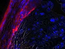
The Chikungunya virus (in red) affects the joints, one of the tissues in which seat the symptoms in humans. /// Copyright M. Lecuit / Institut Pasteur
Sources
“A mouse model for Chikungunya: young age and inefficient type-I interferon signaling are risk factors for severe disease”, PLoS Pathogens, 15 February 2008.
Thérèse Couderc (1,2), Fabrice Chrétien (3,4), Clémentine Schilte (5,6), Olivier Disson (1,2,7), Madly Brigitte (4), Florence Guivel-Benhassine (1,2), Yasmina Touret (8), Georges Barau (8), Nadège Cayet (9), Isabelle Schuffenecker (10), Philippe Desprès (11), Fernando Arenzana-Seisdedos (12,13), Alain Michault (14), Matthew L. Albert (5,6), Marc Lecuit (1,2,7,15)
(1) Microbes and Host Barriers Group, Institut Pasteur, Paris, France
(2) Avenir Team, INSERM U604 Paris, France
(3) Stem Cells and Development Unit, CNRS URA 2578, Institut Pasteur, Paris, France
(4) INSERM, U841, Team 10 Créteil, France; (Public Assistance-Paris Hospitals) Albert Chenevier-Henri Mondor Hospital Group,
Pathology Department, Créteil, France; University of Paris XII, Faculty of Medecine, Créteil, France
(5) Immunobiology and Dendrite Cells Group”, Institut Pasteur, Paris, France
(6) INSERM U818, Paris, France
(7) Bacteria-Cell Interactions Unit, Institut Pasteur, Paris, France
(8) Obstetrics and Gynaecology Department, Reunion South Hospital Group, Saint-Pierre, Reunion Island, France
(9) Ultrastructural Microscopy Platform, Institut Pasteur, Paris, France
(10) National Reference Center for Arboviruses, Institut Pasteur Lyon, France
(11) Flavivirus-Host Molecular Interactions Unit , Institut Pasteur, Paris, France
(12) Molecular Viral Pathogenesis Laboratory, Institut Pasteur, Paris, France
(13) CNRS URA 3015, Paris, France
(14) Laboratory of Microbiology, Reunion South Hospital Group, Saint-Pierre, Reunion Island, France
(15) Necker-Pasteur Centre for Infectious Diseases, Necker-Enfants Malades Hospital (Public Assistance-Paris
Contact persons
Institut Pasteur Press Office :
Marion Doucet - + 33 (0)1 45 68 89 28 - marion.doucet@pasteur.fr
Nadine Peyrolo - + 33 (0)1 45 68 81 47 - nadine.peyrolo@pasteur.fr
Service de presse de l’Inserm
Anne Mignot - + 33 (0)1 44 23 60 73 – presse@inserm.fr


