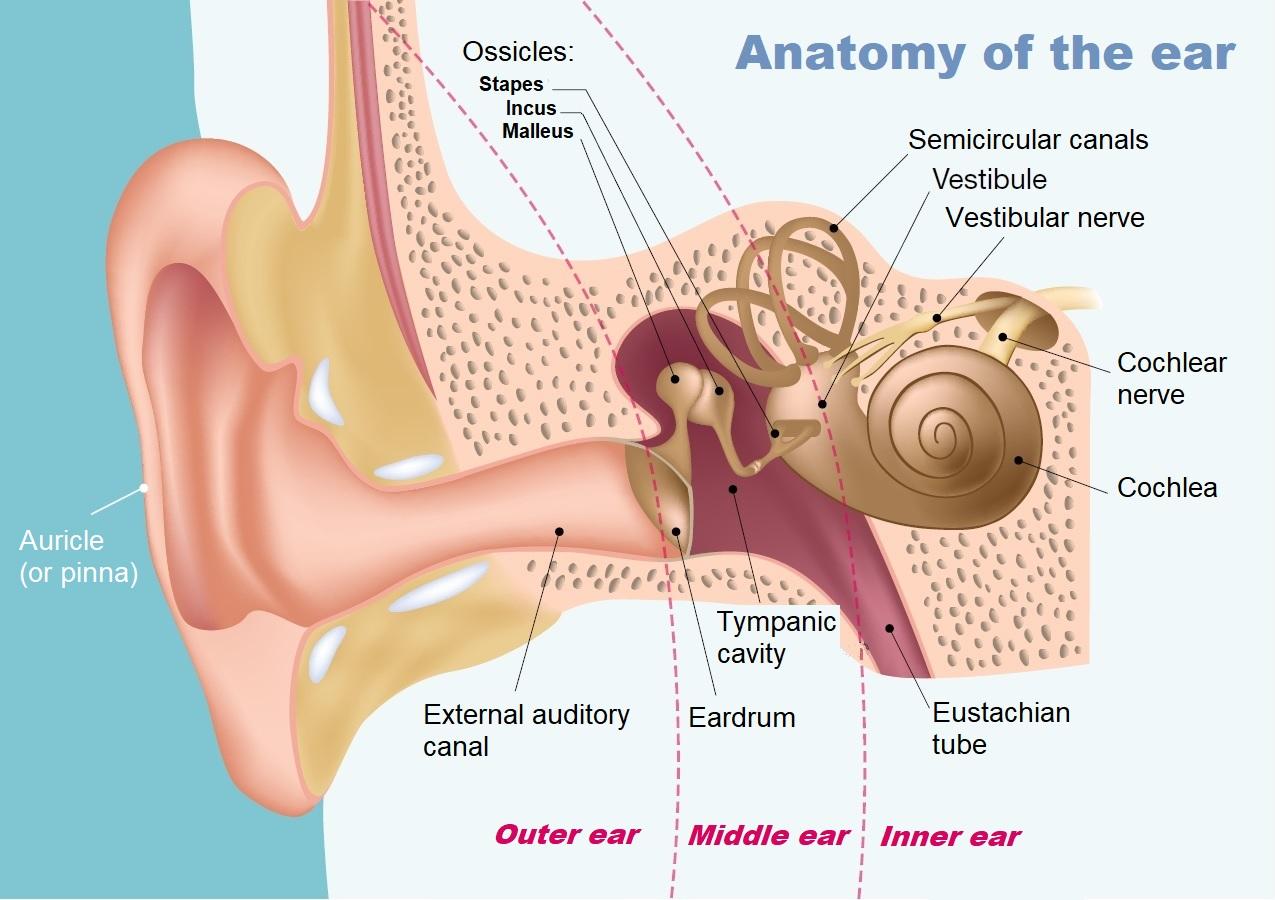
What we generally refer to as the ear is only the visible part. In reality the ear is made up of three parts: the outer, middle and inner ear.
- The outer ear, also known as the auricle or pinna, serves as an acoustic antenna and sensor. Sounds enter the external auditory canal.
- Sound waves then cause the eardrum to vibrate. The eardrum is stretched across the middle ear, which is situated in a bony cavity in the skull and contains three ossicles (tiny bones). The ossicles serve as amplifiers to make up for the loss of energy that occurs when a sound wave travels from the air to the fluid in the cochlea in the inner ear. The malleus picks up the movements of the eardrum and transmits this energy to the incus, which in turn transmits it to the stapes. The stapes (the smallest bone in the human body) is in contact with the membrane covering the "oval window," the entry point into the inner ear, which contains the auditory hearing organ, the cochlea, a sort of long cone composed of three tubes wound into a spiral and filled with fluid.
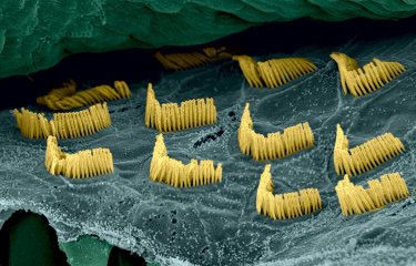
- The central canal houses the auditory sensory organ, the organ of Corti, which is lined with auditory sensory cells known as hair cells (15,000 per cochlea). The hair cells are topped with projections known as stereocilia, which transform sound waves into electrical signals.
- Each sound wave amplified by the middle ear is transformed into a fluid wave, which causes the hair-like stereocilia in the hair cells to vibrate. The hair cells are transducers – they transform the movements of their stereocilia into a nerve impulse that is sent to the auditory nerve. Each of the hair cells lining the cochlea responds preferentially to a given sound frequency (like a set of resonators) so that the brain can differentiate between the pitch of different sounds: high sounds are encoded at the base of the cochlea and low sounds at the apex.
- The brain interprets the signal as a sound at the pitch corresponding to the group of cells stimulated. Sound intensity is analyzed using various mechanisms that depend on the frequency, the rate of discharge of auditory neurons and the nature of the neurons that respond (high- or low-threshold neurons).

The middle ear cavity is linked to the pharynx by the Eustachian tube, whose function is to equalize the pressure on either side of the eardrum – this is very useful during take-off on board a plane, for example. As well as the hearing organ, the inner ear contains the vestibule, the organ that governs the perception of balance by identifying the angle of the head and movements of acceleration. Credits : AdobeStock.


