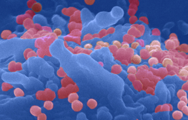
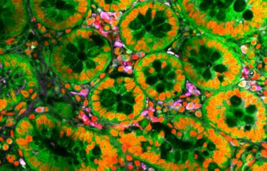
Researchers from Institut Pasteur (Paris), Paris-Descartes University and CNRS, studying HIV-1, have been able to visualize in living cells (direct visualization) the retrotranscribed viral genome, which is capable to integrate into the human genome. They also observed that multiple viral capsid proteins are responsible to lead the retrotranscribed HIV-1 DNA inside the host nucleus. Those direct visualizations are considered a breakthrough in HIV-1 research, and they have been made using the HIV-1 ANCHOR* technology.
*ANCHOR sequences are property of NeoVirTech.
This study entitled “Remodeling of the core leads HIV-1 pre-integration complex in the nucleus of human lymphocytes” addresses a main open question related to the infectious cycle of HIV-1: how the reverse transcribed genome reaches the nucleus. It is important to remember that HIV (Human Immunodeficiency Virus) is an RNA virus. It is part of the family of retroviruses which translates their RNA genome into DNA (retrotranscribed DNA), capable of being integrated into the genome of the host cell.
In the past, the majority of scientific community accepted that a complete core disassembly, a process known as “uncoating”, must take place in the cytoplasm. Answering this question is a difficult challenge because of the typical features that accompany the HIV-1 infection. The details of HIV-1 nuclear translocation process can only be captured by correlative light-microscopy and electron-microscopy.
HIV-1 ANCHOR: a state of the art technology
“To this purpose, we developed a state of the art technology “HIV-1 ANCHOR” that allows for the first time to correlate DNA fluorescence with electron microscopy [correlative light-electron microscopy, CLEM]”, explains Francesca di Nunzio, researcher in the Molecular Virology and Vaccinology Unit, at the Institut Pasteur. “This was so far unattainable due to the incompatibility of fluorescent DNA labelling techniques with EM. Thanks to this novel approach we can now follow the HIV-1 genome in infected cells by live track from the reverse transcription step onward.”
Better understanding of the early steps of HIV-1 infection
This study indicates that the remodeling of cores occurs near the nuclear envelope to give rise the formation of multiple viral capsid (CA) proteins organized in pearl necklace shapes as it has been observed in primary lymphocytes and in HeLa cells.


Viral nuclear complexes at 6 hours post infection contain multiple CA proteins in the nucleus.
First image (on the top): HeLa cells at 6 hours post infection CA gold labelling coupled to transmission electron microscopy forming a pearl necklace-like shape.
Second image (on the bottom): double gold libelling (CA, large dot + DNA, small dot) coupled to transmission electron microscopy HeLa cells at 6 hours post infection. The cartoon on the right represent the viral antigens recognized by the antibodies conjugated with gold particles, such as CA proteins organized in hexamers and/or pentamers and the viral DNA through the ANCHOR system.
Copyright: Institut Pasteur
“Our data argue that the core disassembles prior to nuclear entry into a structure, containing multiple CA proteins, that fits the pore. Thus, our study shed light on critical early steps characterizing HIV-1 infection, thereby revealing novel, therapeutically exploitable points of intervention.” Furthermore, researchers developed and provided a powerful tool enabling direct, specific and high-resolution visualization of intracellular and intranuclear HIV-1 subviral structures.
Live imaging of proviral DNA (green spots in the nucleus), 24 h post infection in HeLa cells.
At the Institut Pasteur, this research was supported Photonic BioImaging Unit (also called Imagopole), which is a technology and service entity providing ultra-structural bio-imaging expertise in life sciences, especially in studies on infectious biology. The study has been carried out by Guillermo Blanco-Rodriguez during his PhD program.
Overall, these findings and technology can be useful for future studies on other pathogens or to investigate the interplay of HIV-1 DNA with nuclear factors and chromatin environments.
This study benefited grants from ANRS and Sidaction.
A patent “HIV-1 ANCHOR technology” is in progress with Institut Pasteur (Paris) in link with NeoVirTech.
Source
Remodeling of the core leads HIV-1 pre-integration complex in the nucleus of human lymphocytes, Journal of Virology, 1st April, 2020, preprint
Guillermo Blanco-Rodriguez 1,2, Anastasia Gazi*3, Blandine Monel*4, Stella Frabetti*1,6, Viviana Scoca*1, Florian Mueller 5,6, Olivier Schwartz 4, Jacomine Krijnse-Locker 3,7, Pierre Charneau 1, Francesca Di Nunzio1
1. Département Virologie, VMV, 28 rue du Dr Roux, 75015 Institut Pasteur, Paris, France
2. École doctorale Frontières de l’innovation en recherche et éducation, CRI, 75004 Paris, université Paris-Descartes, 75006 Paris, France
3. Unité de technologie et de service en bioimagerie ultra-structurale, Centre de ressources et de recherches technologiques, Institut Pasteur, 28 rue du Dr Roux, 75015 Paris, France
4. Département Virologie, UVI, 28 rue du Dr Roux, 75015 Institut Pasteur, Paris, France
5. Département Biologie cellulaire et infection, UIM, Institut Pasteur, 25 rue du Dr Roux, 75015 Paris, France
6. C3BI, USR 3756 IP CNRS, 28 rue du Dr Roux, 75015 Paris, France
7. Paul-Ehrlich-Institut, Langen, Allemagne
This study is part of the priority scientific area Emerging infectious diseases of the Institut Pasteur's strategic plan for 2019-2023.


