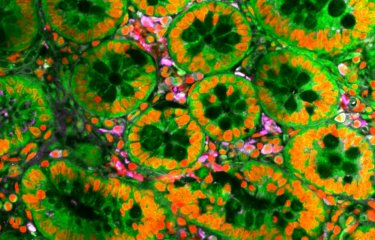
Most of our tissues are constantly renewed and needs to get rid of dying cells without affecting their organisation. A newly identified mechanism is responsible for the seamless exclusion of dying cells from our tissues. This process is based on the dismantling of microtubules, long fibers well known for their function during cell division and transport in the cells. Tuning microtubules stability could be used to exclude cancerous cells from our tissue despite their resistance to cell death.
Epithelia are one of the most common tissues in the human body. Their primary function is to separate organs, maintain their shape and protect them from infections and variations of the environment. These properties are based on the strong adhesions of all cells which seal the tissue layer while maintaining its mechanical cohesion. This stabilising function is however constantly challenged by the turnover of cells, which divide and die very frequently in certain organs. For instance, ten billion of cells are eliminated daily from the human intestine. Cell death is a highly regulated process which is based on the progressive dismantling of cell building-blocks by a family of protein called caspases. Thanks to highly coordinated remodelling processes involving dying cell deformation and shrinkage, those cells can be expulsed from the tissue without impairing its sealing.
A mechanism based on the dismantling of long fibers
However, the connection between the molecular regulator of cell death (the caspases), and the expulsion mechanism remained poorly understood so far. Researchers from the Institut Pasteur (Paris, France), the CNRS, Université Paris Cité and Sorbonne Université found a new central player of this process which is based on the dismantling of long fibers, called microtubules, by caspases. This work has been published in Nature Communications in June 2022.
This surprising observation was performed in the thorax epithelium of the fruitfly Drosophila. “This little fly is an ideal system to understand the mechanisms of dying-cell exclusion: the regulators of cell death are similar to the one in humans and the organisation of epithelia is very similar. Moreover, living cells and tissues can be easily imaged on microscopes while using the numerous genetic tools available in flies to perturb the process” say Dr Romain Levayer, corresponding author of this study.
Observing the molecular regulators required to deform cells
Using highly resolutive imaging of living tissue in the fly pupae (the “chrysalid” stage of the fly), the authors first found that the initial deformations of the dying cells were not driven by the usual factors orchestrating cell deformation, the actin filaments and the molecular motor Myosin II. “We knew a lot about the molecular regulators required to deform cells. This is usually driven by a little molecular motor named Myosin II bound to filaments called actins which can contract cells just like little muscles” said Alexis Villars, first author of this study. “We could not observe any modification of these motors despite the rapid deformations of the dying cells preceding their expulsion, suggesting that another unknown process was responsible for these initial deformations”.
The role of microtubules in the regulation of cell shape
The answer emerged when the authors looked at another component of the cell architecture: the microtubules. Microtubules are dynamic hollowed tubes which span the entire cells and form efficient highway to transport components across the cells. Microtubules are also well known for their role in cell division and the repartition of the genetic information between the two daughter cells. However, their architectural roles remained poorly studied so far. “The initiation of cell expulsion was perfectly concomitant with the dismantling of microtubules throughout the dying cell by caspases. This was suggesting that they could play a central role in this process” says Alexis Villars.
Bypassing the needs for caspases to expulse cells
Accordingly, stabilising microtubules prevented dying cell expulsion, while genetic and chemical disassembly of microtubules were sufficient to expulse cells from the tissue, even when perfectly healthy or when caspases were completely blocked. “This was a very important result, as it is showing for the first time that we could bypass the needs for caspases to expulse cells” says Romain Levayer.
“These results also illustrate the underestimated role of microtubules in the regulation of cell shape, which like iron rods embedded in concrete, can stabilise the shape of our cells”. Cancerous cells are frequently protected from death thanks to genetic mutations altering caspase activity. The modulation of microtubules with drugs could help to expulse these abnormal cells from our tissue despite their resistance to cell death.
This study is part of the Cancer Initiative of the Institut Pasteur's strategic plan for 2019-2023.
Source
Microtubules depletion by effector caspases is an important rate-limiting step of extrusion, Nature Communications, June 25, 2022.
doi: 10.1038/s41467-022-31266-8
Alexis Villars1,2, Alexis Matamoro-Vidal1, Florence Levillayer1 and Romain Levayer1
1. Department of Developmental and Stem Cell Biology, Institut Pasteur, Université Paris Cité, CNRS UMR 3738, 25 rue du Dr. Roux, 75015 Paris
2. Sorbonne Université, Collège Doctoral, F75005 Paris, France


